


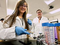


What's the biggest strength of the department?
Our strength is that we have a wide diversity of perspectives under one roof: we have cell biologists, biochemists, molecular biologists, geneticists and structural biologists. They can attack the same problem at different scales and in different ways. It gives a much broader understanding of a problem.
How do you see the department growing or changing in the next few years?
One big area of growth is structural biology, particularly cryo-electron microscopy, which is revolutionizing biology. We are just finishing construction on a new state-of-the-art cryo-EM facility, which will enhance many of our research programs. We will also start to collaborate with UT Austin's new Dell Medical School. This will enrich our work by allowing us to go from model systems into disease systems, by giving us access to clinical samples and by exposing us to ideas and problems that may benefit from our basic-science perspective.
What's the biggest challenge facing the department?
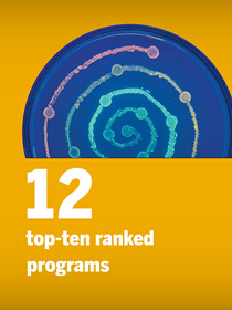
Our reputation hasn't caught up to the caliber of our work. My vision is that when you ask people to name the top two or three molecular biology programs in the country, along with the ones mentioned today, they would say The University of Texas at Austin.
How do you get there?
We'll continue to hire world-class faculty, build state-of-the art facilities and lead in teaching. We've innovated in science education with programs like the Freshman Research Initiative and UTeach, and we have a culture that values teaching. That enhances our scholarship at all levels and helps us attract and train the best students. Remaining on the cutting-edge of science education is essential to staying relevant and moving forward in the future.
You've just established the Sauer Center for Structural Biology, a facility for taking 3D snapshots of molecules with cryo-EM. What kinds of research will this new facility enable?
Virtually any biochemical process — from copying DNA to building proteins to turning light into metabolism through photosynthesis — involves molecules behaving and moving. And we want to see how those processes work. That can help us design better drugs, understand the causes of diseases and make better crops. It's not a guarantee, but the more you understand at a molecular level, the more insight you can have to use biology to make human lives better.
Why is this technique so powerful?
Cryo-EM allows you to take pictures of proteins and other molecules at the resolution of atoms. With X-ray crystallography, an older way of getting atomic resolution structures, you have to grow crystals out of the molecules and that's hard to do for large molecules. But with cryo-EM, the bigger the molecule the better. So they're complementary methods. Cryo-EM enables us to look at things that we didn't have a prayer of seeing at atomic resolution with prior ways. It also allows us to see the different shapes that a molecule takes, such as the active and inactive states of cell receptors. That is also harder to do with other methods.
Why is it useful to see molecules in such detail?
In structural biology, seeing is believing. A picture of a molecule helps us figure out how it works and how it interacts with other molecules to create living cells and tissues. It's like taking apart a clock and studying all of the pieces to understand how it works.
What kinds of molecules or processes will you personally be studying with cryo-EM?
I study epidermal growth factor, an important receptor on cell surfaces involved in development and in healing skin after a wound. Tickle this receptor and the cells grow and divide, so mutated versions of the receptors are often involved in cancer. Consequently, many cancer drugs target these receptors. In my work, I try to see how the receptors and the drugs that target them work.
You played a role in getting a new breast cancer treatment into clinical use. Can you tell me about that?
A particularly aggressive form of breast cancer is caused by overexpression of a receptor called HER2. There was already a drug on the market called Herceptin that targeted HER2, but it didn't work in some people, and in others, tumors often evolved resistance. Genentech had discovered another drug called Pertuzumab that also targeted HER2. We could see that the two drugs bound to different parts of HER2 and would inhibit its function in different ways. We suspected that combining the two drugs would have a synergistic effect. On the basis of this and other observations, Genentech decided to push Pertuzumab through clinical trials and received approval to market it. Now these two drugs are used together and, just as we suspected, they are more effective than Herceptin alone. I'm very proud of this work but wish to emphasize that our contribution was one piece of a large puzzle. Also, Gail Lewis, a UT alumna and scientist at Genentech, played key roles in the development of Herceptin and Pertuzumab.


Comments